Blog Template Not Full Width

An oocyte is the very beginning of human life. In simple terms, it is an immature egg cell.
Throughout the process of ovulation, this immature egg cell eventually matures and becomes an ovum or egg.
Spindle Imaging allows embryologists to check for critical structures in the human oocyte.
In other words, spindle imaging uses a powerful polarized microscope to check the human oocyte for any abnormalities.
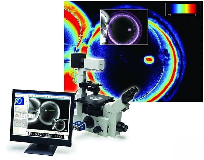
Some oocyte may look normal but upon inspection with spindle imaging, an embryologist can detect the various abnormalities or defects like missing or misplaced spindle and polar body malformations.
The spindle is where the DNA is located.
It is the structure that carries the DNA of the egg.
A matured egg when it is ejected from the ovum should have the presence of a spindle in the correct location beneath the polar body.
If the spindle could not be seen under a polarized microscope, that means the quality of the egg is extremely poor.
An egg may have a spindle but the spindle may be in a different location!
All the eggs that have a spindle that is drifted too far away from the polar body has a high chance of being abnormal.
This means that the egg has a high chance of genetic abnormalities.
An embryologist will orientate the egg to ensure the location of the spindle is at 6 or 12 o' clock position during the microscope injection process.
All these abnormalities cannot be detected with a normal microscope.
It can only be detected using spindle imaging technology.
The aim of the embryologist is to inject good quality sperm into a good quality egg for implantation.
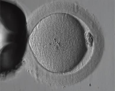
Conventional Contrast Image
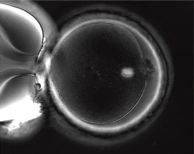
Spindle image where spindle is seen clearly
In a conventional contrast image (left) of a human oocyte taken just prior to ICSI, structures such as the spindle and multiple layers of the zona pellucida remain invisible.
With spindle imaging (right) the spindle is clearly seen to be nicely barrel shaped and the three layers of the zona pellucida are all visible.
Key Benefits
Unprecedented Resolution
High-contrast live images of the oocyte and spindle
Non-invasive Imaging
Does not require the use of any labels or stains, preserving the biology of the spindle and related structures
Quantitative Analysis
Tracks oocyte behaviour over time and automatically records data points of molecular density and orientation
If you would like to know more about spindle imaging technology and how it is related to embryo development in IVF treatment, call or whatsapp our IVF Penang centre to start your fertility journey with us.